
Dr. Christine P. Hendon: "Fully Automated Postlumpectomy Breast Margin Assessment Utilizing Convolutional Neural Network Based Optical Coherence Tomography Image Classification Method"
Date: October 18th, 2020 - See the recording of our event below!
For this interview, we will be discussing her paper “Fully Automated Postlumpectomy Breast Margin Assessment Utilizing Convolutional Neural Network Based Optical Coherence Tomography Image Classification Method”, in which she discusses a way to analyze optical coherence tomography (OCT) images of breast tissue using the Convolutional Neural Network and optimize cancer region classification in those images.
PAPER SUMMARY
Key Terms:
- (OCT) optical coherence tomography - a high-speed, microscopic imaging modality. It is similar to ultrasound, using light instead of sound to produce micron-scale resolution
- (CNN) Convolutional Neural Network - the algorithm used to classify cancer in OCT images of breast tissue.
- Lumpectomy - a surgery to remove breast tissue (only the tumor and a small ring of normal tissue around it.
Breast cancer is generally detected through a doctor’s examination of imaging or screening. Current methods of manual interpretation of breast cancer imaging leave room for error, and about 23% of patients end up having to have multiple surgical removals due to this.
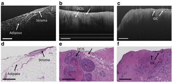
Based on OCT images of cancerous and non-cancerous breast tissue, a CNN algorithm was developed to automate detection of cancer. 46 samples were used, with 4 different types of tissue, including adipose (fatty tissue), stroma (connective tissue), ductal carcinoma in situ (cancerous @ original site), and invasive ductal carcinoma (cancerous & migrating elsewhere). The algorithm’s classification of each sample was confirmed through histology, or tissue staining and manual examination.
The CNN algorithm turned out to be able to differentiate between normal, healthy tissue, and cancerous tissue. It worked much more quickly than manual analysis, and various specialists were able to be trained to understand the images after an average of 3.4 hours of training, suggesting that this is a very realistic and user-friendly option. On the other hand, the study was conducted on a fairly small sample size, thus many more samples would need to be incorporated to develop its accuracy and adaptability. Histology of these samples would need to be accurate as well, or it can potentially train the algorithm to incorrectly differentiate between samples.
OCT images of breast tissue structures acquired with ultrahigh-resolution OCT (UHR-OCT) system
(a) Stroma and adipose
(b) Ductal carcinoma in situ (DCIS)
(c) Invasive ductal
carcinoma (IDC)
(d-f) Images of corresponding histology
Read Dr. Hendon's paper here